CT扫描方案
CT扫描方案
PG电子APP的3D打印和培训中心, we are dedicated to providing cutting-edge solutions for medical professionals and 研究员s. 我们的CT扫描方案确保了医学成像的最高质量和精度. 该协议是一个全面的指南,可以帮助您浏览该过程, 从数据采集到3D建模. Explore our detailed CT扫描方案 below to optimize your experience and access the valuable resources we offer. 无论您是医疗保健提供者, 研究员, 或教育家, 我们最先进的技术和专业知识随时为您的需求提供支持.
感谢您抽出时间阅读本协议. The quality of the CT scan is the most important aspect of creating case- specific anatomical models. Your observation of the recommendations made in this protocol will have a significant impact on the accuracy of the final model. We understand concerns about keeping the 辐射剂量 to your patients as low as reasonably achievable, 因此, 请适当地将这些指导原则应用到你的病人身上. Please do not hesitate to contact us toll free at (402)552-3569 with any questions or prior to using this protocol for the first time.
请牢记以下要点:
- Please use a 3D scanning routine that provides high resolution images as would be suitable for image guided surgery, 立体规划或其他3D应用. 咨询您的CT供应商可能会有所帮助 Application Specialist for advice on optimal parameters for your machine that provide the best scan with acceptable 辐射剂量 levels. 扫描不应在手术日期前超过6个月.
- 获取高空间分辨率的扫描. 系列 should be acquired with thin, contiguous image slices (equivalent thickness and spacing of 1.25 mm or less) and as small a field of view (FOV) as possible while still including the patient’s anatomy of interest.
- 请提供原始扫描平面的图像. 如果执行软件后处理以重新定向或重新格式化扫描卷, 那么必须包含原始采集平面的一系列薄切片图像.
- 在图像采集过程中不使用龙门架倾斜. Images acquired with gantry tilt then post-processed to reorient images are not acceptable (i.e. “取出”倾斜) .
- 请确保扫描没有运动伪影. 在整个扫描过程中,病人必须保持完全静止. 如果患者出现运动,则必须重新启动扫描. 患者运动引起的图像失真会严重损害模型的准确性.
- 金属植入物引起的图像伪影会模糊感兴趣的解剖结构. 请采取措施尽量减少金属制品的存在.
- 存档整个研究在未压缩的DICOM格式的CD-R或DVD运输. 通过互联网传输图像数据也是可能的. 详情请致电(402)552-3569查询.
医用CT扫描仪的推荐方案
| 病人定位: | 仰卧的 |
| 片间距 | 1.25mm以下(等于切片厚度) |
| 像素大小: | 0.60毫米或以下 |
| 视野: | 设置视野以包括整个感兴趣的区域 |
| 算法(示例): |
GE-标准(非骨或细节) |
| 龙门倾斜: | 0 o |
| 存档介质: | CD或DVD |
| 文件类型: | DICOM(压缩) |
| 系列 |
原始/主/轴(无重构、重新格式化或处理数据) |
感谢您抽出时间阅读本协议. The quality of the CT scan is the most important aspect of creating case- specific anatomical models for bolus production. Your observation of the recommendations made in this protocol will have a significant impact on the accuracy of the final bolus. We understand concerns about keeping the 辐射剂量 to your patients as low as reasonably achievable, 因此, 请适当地将这些指导原则应用到你的病人身上. Please do not hesitate to contact us toll free at (402)552-3569 with any questions or prior to using this protocol for the first time.
- Please use a 3D scanning routine that provides high resolution images as would be suitable for image guided surgery, 立体规划或其他3D应用. 咨询您的CT供应商可能会有所帮助
Application Specialist for advice on optimal parameters for your machine that provide the best scan with acceptable 辐射剂量 levels. 扫描不应在第一次治疗日期前超过1个月进行.
- 获取高空间分辨率的扫描. 系列应与薄获得, contiguous image slices (equivalent thickness and spacing of 3 mm or less) and as small a field of view (FOV) as possible while still including the patient’s anatomy of interest.
- 请提供原始扫描平面的图像. 如果执行软件后处理以重新定向或重新格式化扫描卷, 那么必须包含原始采集平面的一系列薄切片图像.
- 如果蛀牙(e).g. 内耳、外耳、鼻子等.) will be packed during treatment, acquire the scan with tissue-equivalent packing material. 另外, contour packing material during the planning process prior to DICOM submission to 3D Systems.
- 在图像采集过程中不使用龙门架倾斜. 使用龙门倾斜获取图像,然后进行后处理以重新定位图像(i.e. “取出”倾斜)是不可接受的.
- 请确保扫描没有运动伪影. 在整个扫描过程中,病人必须保持完全静止. 如果患者出现运动,则必须重新启动扫描. 患者运动引起的图像失真会严重损害模型的准确性.
- 金属植入物引起的图像伪影会模糊感兴趣的解剖结构. 请采取措施尽量减少金属制品的存在.
- 存档整个研究在未压缩的DICOM格式的CD-R或DVD运输. 通过互联网传输图像数据也是可能的. 详情请致电(844)643 1001与我们联系.
医用CT扫描仪的推荐方案
| 片间距: | 3毫米或以下(等于切片厚度) |
| 千伏峰值: | 90 - 140 |
| 视野: | 设置视野以包括整个感兴趣的区域 |
| 算法(示例): |
GE-标准(非骨或细节) |
| 龙门倾斜: | 0 o |
| 存档介质: | CD或DVD |
| 文件类型: | DICOM(压缩) |
| 系列: | 原始/主/轴(无重建、重新格式化或后处理数据) |
The quality of the CT scan is the most important aspect of creating a patient matched surgical plan. The purpose of this protocol is to obtain patient imaging data suitable for 3D reconstruction of the anatomy. Your observation of the recommendations made in this protocol will have a significant impact on the accuracy of the models, 指南, 以及为病人创建的模板. We understand concerns about keeping the 辐射剂量 to your patients as low as reasonably achievable, 因此, 请适当地将这些指导原则应用到你的病人身上. Do not hesitate to contact us toll free at (844) 643-1001 with any questions prior to using this protocol.
- Please use a 3D scanning routine that provides high resolution images as would be suitable for image guided surgery, 立体定向计划, 或其他3D应用程序. It may be useful to consult with your CT vendor’s Application Specialist for advice on optimal parameters for your machine that provide the best scan with acceptable 辐射剂量 levels. Scans should not be taken more than 6 months prior to surgery date for patient 21 years of age or older.
- 序列应该用薄的、连续的图像切片(等效厚度为1.25mm or less) and as small a field of view as possible while still including the patient’s anatomy of interest.
- 请提供原始扫描平面的图像. 如果执行软件后处理以重新定向或重新格式化扫描卷, 然后必须包含原采集平面的一系列薄切片图像.
- 在图像采集过程中不使用龙门架倾斜. 通过龙门倾斜获取的图像,然后进行后期处理以重新定位图像是不可接受的.
- 确保扫描没有运动伪影. 在整个扫描过程中,病人必须保持完全静止. 如果患者出现运动,则必须重新启动扫描. 患者运动引起的图像失真会影响准确性.
- 金属植入物引起的图像伪影会模糊感兴趣的解剖结构. Please take steps to minimize metal artifacts; including artifact caused by contralateral implants. Elevation of the contralateral limb to reduce metal artifact scattering to the anatomy of interest is 首选.
- 存档整个研究在未压缩的DICOM格式的CD-R或DVD运输. 通过互联网传输图像数据也是可能的. 详情请致电(844)643-1001与我们联系.
扫描参数:
| 病人定位: | 使患者处于舒适、稳定的仰卧位. 为使患者感到舒适,可以采用其他体位, 完整视野, 最小化 辐射剂量. |
| 片间距: | 1.25mm以下(等于切片厚度) |
| 矩阵: | 512 X 512 |
| 千伏峰值: | 90 - 140 |
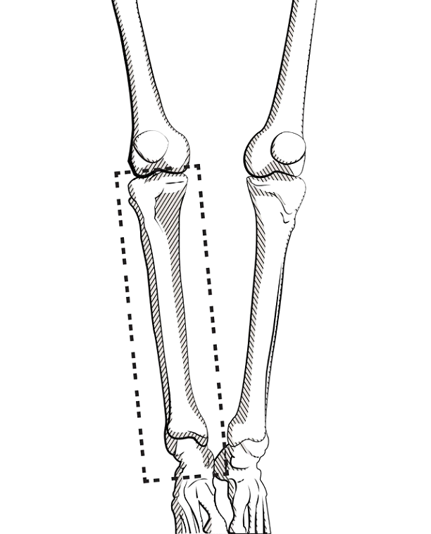
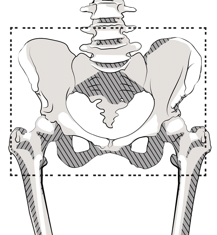
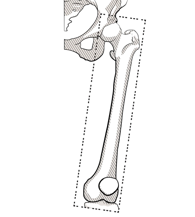
| 视野: | Capture entire bony segment of interest from joint to joint using smallest field of view possible. 请看下面的例子. |
| 算法(示例): | 标准软组织重建算法 首选. GE:标准(非骨或细节) 西门子:H30s 东芝:FC20 飞利浦:B |
| 龙门倾斜: | 0 o |
|
存档介质: |
CD或DVD |
| 文件类型: | DICOM(压缩) |
| 系列: | 原始/主/轴(无重建或后处理数据) |
盆腔字段:
股:
胫骨字段: